ROI-Guided Lumbar Spine Degeneration Detection

Lumbar Spine Degeneration, otherwise known as Disk Degeneration Disease, is the deterioration/weakening of the intervertebral disks in the lower back, causing them to lose their ability to absorb shock and potentially leading to pain and discomfort due to nerve compression. While lumbar degeneration is a natural part of aging, its progression and severity can vary from person to person. Early detection and classification of the type and stage of degeneration are crucial for effective management and treatment. We are using the power of AI to automate the detection of lumbar degeneration.

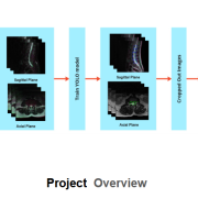
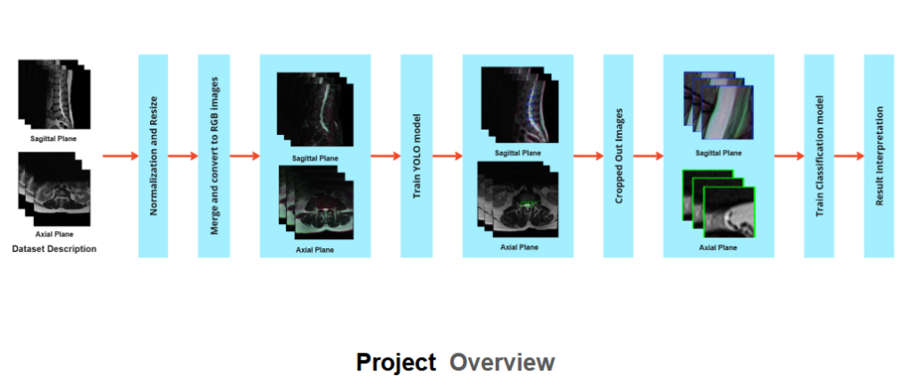
Firstly, We merge and pre-process the Magnetic Resonance Imaging (MRI) scans to convert them to RGB images, comprised with multi-label diseases per image.Then, we used an YOLO(You Only Look Once) architecture to detect the region of interests (ROI) that include the intervertebral disks in the MRI scans from both Sagittal and Axial Plane. Finally, the ROIs are classified into different degree of degeneration.

The goal of this research is to detect Lumbar Spine Degeneration as early as possible in order to ensure the best quality of life and outcome for the patient.

Detection-Guided Kidney Abnormality Segmentation

Tumors and cysts are two major kidney abnormalities which can lead to cancer if left undetected and untreated timely. The current diagnostic process depend on computed tomography (CT) scan screening which is time-consuming and specialist-dependent. This leads to fatigue for radiologists and doctors. As a result, diagnostic errors increase. To enhance the diagnostic process, we are developing an AI-based automated method for kidney abnormality detection. 

The first step in this method is kidney detection using the YOLOv8 model. This is conducted on 2D sliced images extracted from 3D CT. The detected kidney regions are then cropped to reduce the background area. After that, abnormal region segmentation is conducted using the U-Net segmentation model. The abnormal region consists of either cyst or tumor or both. This produces a mask of abnormal region for each 2D slice. The 2D mask slices are combined to construct a 3D mask of the abnormal region. 

This study aims to automate the detection process of kidney tumor and cyst, offering a faster and enhanced approach to assist the doctors and radiologists in diagnostic process.

Relevant publications:

1. J. Faruk, S. B. Alam, S. S. T. Elma, S. Wasi, R. Rahman and S. Kobashi, “Kidney Abnormality Detection Using Segmentation-Guided Classification on Computed Tomography Images,” 2024 International Conference on Machine Learning and Cybernetics (ICMLC), Miyazaki, Japan, 2024, pp. 414-419, doi: 10.1109/ICMLC63072.2024.10935113.

2. S. Wasi, S. B. Alam, R. Rahman, M. A. Amin and S. Kobashi, “Kidney Tumor Recognition from Abdominal CT Images using Transfer Learning,” 2023 IEEE 53rd International Symposium on Multiple Valued Logic (ISMVL), Matsue, Japan, 2023, pp. 54-58, doi: 10.1109/ISMVL57333.2023.00021

Analyzing rice genomic variation along with expression and phenotype data to reveal stress tolerance mechanism focusing development of climate smart rice